Bonevia
The 2026 Best Craniofacial Implants Manufacturers Worldwide guide examines companies shaping modern facial reconstruction. Craniofacial Implants can restore contour, protect exposed bone, and support functional recovery. Their value depends on more than attractive product photographs. It depends on clinical evidence, material performance, surgical compatibility, and consistent manufacturing.
Dr. Paul Manson, a respected craniofacial surgeon, has emphasized, “Craniofacial surgery is a team effort.” This principle remains highly relevant. Surgeons, biomedical engineers, radiologists, and manufacturers must work together before implantation begins. A patient-specific implant may start with thin-slice CT data. Engineers then create a digital model, check symmetry, and select titanium, PEEK, or another suitable material. The final device should fit the patient’s anatomy, not merely match a catalogue size.
This overview considers design capability, customization, quality systems, regulatory documentation, production precision, and after-sales support. It also looks at experience with trauma, congenital differences, tumor reconstruction, and revision procedures. Reliable manufacturers should explain sterilization methods, traceability, material testing, and clinical limitations clearly. Marketing language alone is not enough.
No ranking is perfect. That limitation matters. Manufacturer performance can vary by region, application, and hospital partnership. Buyers should verify current certifications, published evidence, surgeon feedback, and local approval requirements. A lower price may conceal design compromises or weak support. A premium price may not guarantee better outcomes. Careful comparison remains essential when selecting Craniofacial Implants for complex clinical needs.
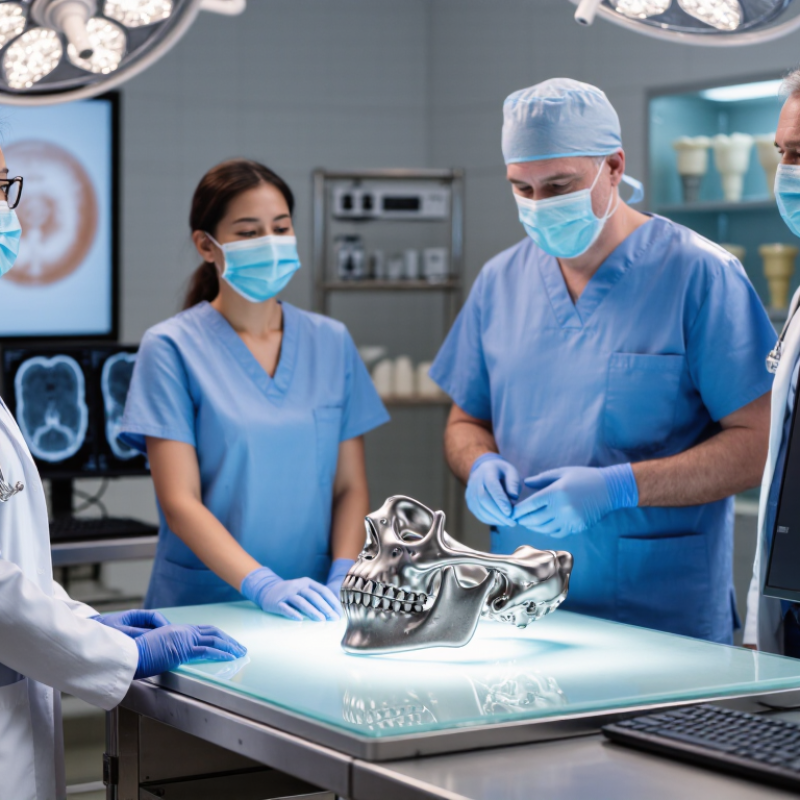
Craniofacial implants restore form and function after trauma, tumor removal, infection, or congenital differences. They may protect exposed bone, support soft tissue, and improve facial symmetry. In practice, the purpose is not purely cosmetic. A well-planned implant can help protect the brain, rebuild an orbital wall, or restore chewing and breathing pathways.
Common types include cranial plates, orbital implants, midface implants, nasal supports, and auricular frameworks. Surgeons may select titanium, porous polyethylene, PEEK, or other medically approved materials. Patient-specific implants are designed from CT data and can match irregular bone defects closely. Standard implants may suit simpler repairs and reduce planning time. Small details matter. A few millimeters can affect eye position, occlusion, or facial balance.
Clinical application requires more than accurate imaging. A craniofacial team reviews bone quality, infection risk, soft-tissue coverage, and the patient’s long-term needs. Surgeons, radiologists, engineers, and reconstructive specialists often share the plan. Follow-up checks healing, implant stability, sensation, and appearance. Complications can include infection, exposure, migration, or poor integration. No implant is perfect. Even strong digital planning can miss tissue changes or patient expectations. That limitation deserves honest discussion before surgery. Selection should follow validated clinical evidence, careful manufacturing controls, and applicable medical-device requirements.
Modern craniofacial implant manufacturing begins with accurate patient data. High-resolution CT scans capture bone defects in three dimensions. Engineers convert these images into digital anatomical models using specialized CAD software. Surgeons can then review implant contours before production begins.
Fit matters.
Computer numerical control machining supports precise work with titanium and high-performance polymers. Additive manufacturing can create complex lattice structures that reduce weight and support tissue integration. Porous surfaces may improve fixation, but their performance depends on pore size, strength, and clinical use. Material selection remains a careful balance between durability, imaging compatibility, and biological response.
Quality systems connect every stage, from design verification to dimensional inspection. Optical scanning and coordinate measurement machines detect small contour errors. Some facilities use surgical navigation data to refine implant positioning. Production teams must also document cleaning, packaging, sterilization, and traceability according to applicable medical-device requirements. Digital workflows improve consistency, yet they do not remove clinical judgment. A beautiful digital model can still fail if the scan is incomplete or the patient’s anatomy changes.
Experienced manufacturers therefore combine engineering review with surgeon feedback and case-specific planning. They test mechanical behavior, inspect surface quality, and record deviations honestly.
No process is flawless. Continuous review is essential.
When evaluating craniofacial implant manufacturers, start with clinical experience, not polished brochures. Ask how long they have produced patient-specific and standard implants. Review case histories involving orbital, cranial, and facial reconstruction. Details matter. Speak with surgeons who have handled the implants in real operating rooms. Ask about fit, contour accuracy, handling, and unexpected revisions. A manufacturer’s experience should appear in documented outcomes, not only sales language.
Examine the technical evidence behind each product. Check material specifications, imaging compatibility, design tolerances, and validated manufacturing controls. Request information about sterilization, packaging, batch traceability, and complaint handling. A dependable supplier should explain how digital files become a finished implant. They should also show how engineers verify margins, screw placement, and anatomical symmetry. Ask for evidence. If answers remain vague, the evaluation is incomplete.
Quality systems deserve direct attention. Look for current certifications, documented risk management, trained personnel, and consistent inspection records. Regulatory status should be verified for the intended market and implant category. Response time matters when anatomy changes or a case becomes urgent. Test communication with a sample planning request. Notice whether questions receive precise answers or generic promises. I would not ignore cost, but price alone can mislead. A lower quote may exclude planning support or revision assistance. One weakness in this process is relying too heavily on paperwork. Site audits, surgeon feedback, and follow-up data can reveal what documents miss. Manufacturers should welcome that scrutiny.
Leading Craniofacial Implant Manufacturers Worldwide in 2026 are judged by more than production scale. Clinical reliability, material knowledge, and documented quality systems matter equally. Experienced manufacturers work closely with craniofacial surgeons, biomedical engineers, and hospital procurement teams. This cooperation helps translate complex anatomy into safer implant designs.
Strong suppliers usually offer patient-specific solutions supported by high-resolution imaging and precise digital planning. Their manufacturing processes should control materials such as titanium, PEEK, or medical-grade polymers. Each stage needs clear traceability, from raw material inspection to final packaging. Sterilization guidance and storage instructions should be practical for hospital staff. Small details matter.
Not every impressive catalogue proves clinical excellence. Public evidence can be incomplete. Buyers should examine validation reports, post-market feedback, and technical support records. They should also ask how manufacturers handle design changes and unexpected fit problems. A responsive engineering team can reduce delays during difficult reconstructions. However, customization may increase costs, planning time, and review requirements. The best choice depends on anatomy, surgical experience, hospital resources, and local regulatory expectations. Some manufacturers still communicate these limitations poorly. That deserves closer scrutiny.
| Implant Category | Common Clinical Applications | Typical Materials | Key Design Characteristics | Manufacturing Technologies | Clinical and Regulatory Considerations |
|---|---|---|---|---|---|
| Cranial Reconstruction Implants | Repair of cranial defects following trauma, decompressive craniectomy, tumor resection, or congenital abnormalities. | PEEK Titanium Porous polyethylene PMMA | Patient-specific contour matching, fixation holes, edge transition control, anatomical symmetry, and compatibility with postoperative imaging. | CT-based CAD/CAM, additive manufacturing, CNC machining, molding, polishing, and inspection against digital anatomical models. | Implant design must support stable fixation and soft-tissue coverage. Material selection should consider strength, radiographic visibility, infection risk, and revision requirements. |
| Orbital Floor and Midface Implants | Reconstruction of orbital floor defects, zygomaticomaxillary injuries, and selected midface contour deficiencies. | Titanium mesh PEEK Porous polyethylene | Thin profile, anatomical curvature, controlled flexibility, smooth tissue-facing surfaces, and secure fixation points. | Laser-cut or additively manufactured metal structures, thermoforming, CNC machining, and patient-specific digital planning. | Critical considerations include orbital volume restoration, avoidance of soft-tissue entrapment, implant stability, and verification of anatomical fit by imaging. |
| Facial Contour Implants | Reconstruction or augmentation of the chin, cheek, malar region, forehead, and other facial contours. | Silicone elastomer PEEK Porous polyethylene Titanium | Contoured geometry, low-profile margins, tissue compatibility, fixation features, and options for stock or patient-specific designs. | Compression molding, injection molding, CNC machining, additive manufacturing, and silicone finishing processes. | Product evaluation generally addresses biocompatibility, mechanical integrity, dimensional accuracy, sterilization compatibility, and long-term soft-tissue interaction. |
| Temporomandibular Joint Components | Reconstruction of the mandibular condyle and glenoid fossa in severe joint disease, trauma, ankylosis, or revision cases. | Cobalt-chromium alloys Titanium alloys Ultra-high-molecular-weight polyethylene | Articulating bearing surfaces, modular components, fixation screws, anatomical alignment, and resistance to cyclic loading. | Investment casting, CNC machining, additive manufacturing, surface finishing, and precision assembly. | Design assessment typically focuses on wear, fatigue strength, fixation stability, range of motion, dislocation risk, and component compatibility. |
| Mandibular Reconstruction Implants | Segmental or partial mandibular reconstruction after trauma, tumor surgery, osteonecrosis, or major bone loss. | Titanium alloys PEEK Titanium plates | Load-bearing geometry, patient-specific curvature, screw-hole positioning, bone-graft integration space, and occlusal alignment support. | Patient-specific additive manufacturing, CNC machining, bending and forming, laser cutting, and digital surgical planning. | Important factors include fatigue resistance, fixation strength, fit accuracy, interaction with bone grafts, and compatibility with reconstructive surgical workflows. |
| Maxillofacial Fixation Systems | Stabilization of facial fractures and osteotomies involving the maxilla, mandible, orbit, zygoma, and cranial facial skeleton. | Titanium Stainless steel Bioresorbable polymers | Low-profile plates, locking or non-locking screws, multiple plate geometries, controlled stiffness, and intraoperative contourability. | Precision stamping, laser cutting, CNC machining, injection molding, passivation, cleaning, and validated sterilization packaging. | Performance requirements include screw-plate compatibility, corrosion resistance, mechanical stability, sterilization assurance, and appropriate radiographic assessment. |
| Patient-Specific Craniofacial Implants | Complex anatomical defects where standard implants cannot provide adequate fit or symmetry. | PEEK Titanium PMMA Porous polyethylene | Customized from medical imaging, with individualized margins, fixation features, thickness, curvature, and anatomical landmarks. | Digital segmentation, virtual surgical planning, CAD/CAM, metal or polymer additive manufacturing, and dimensional quality inspection. | Manufacturers must control image-to-design traceability, software validation, design verification, manufacturing reproducibility, and patient-specific production records. |
| Porous and Osteoconductive Designs | Applications where bone ingrowth, biological fixation, or enhanced soft-tissue integration is a design objective. | Porous titanium Hydroxyapatite-coated materials Porous polyethylene | Controlled pore architecture, interconnected porosity, optimized surface roughness, and balance between mechanical strength and tissue integration. | Metal additive manufacturing, surface treatment, coating deposition, pore-structure inspection, and cleaning validation. | Evaluation should address pore consistency, coating adhesion where applicable, particulate control, sterilization, biocompatibility, and mechanical performance. |
| Quality and Compliance Capabilities | Applies across craniofacial implant categories and supports consistent design, production, inspection, and post-market monitoring. | Implant-grade metals Medical polymers Ceramic coatings | Documented design controls, risk management, traceability, validated processes, nonconformance control, and change management. | Coordinate measuring machines, optical scanning, CT inspection, surface metrology, mechanical testing, clean manufacturing, and validated sterilization processes. | Common reference frameworks include ISO 13485 for quality management, ISO 14971 for medical-device risk management, ISO 10993 for biological evaluation, and applicable regional medical-device regulations. |
| Technical requirements, regulatory classification, labeling, and clinical evidence vary according to the implant’s intended use, anatomical location, material, design, and jurisdiction. The table summarizes widely used industry dimensions without ranking or identifying individual companies or brands. | |||||
2026 Best Craniofacial Implants Manufacturers Worldwide
Safety now defines leadership in craniofacial implant manufacturing. The World Health Organization reports that approximately one in ten patients experiences harm during healthcare delivery. Implant planning must therefore extend beyond shape and surgical fit. Manufacturers should document material traceability, sterilization validation, fatigue testing, and post-market surveillance. ISO 10993-1 remains important for biological evaluation, while ISO 13485 supports controlled quality systems. In the United States, regulatory submissions require evidence that matches the device’s risk profile. Under the European Union Medical Device Regulation, clinical evaluation and lifetime monitoring receive greater attention. Compliance is not paperwork. It protects bone, tissue, and trust.
Market forecasts remain inconsistent. Reports from Grand View Research and MarketsandMarkets project steady growth in craniofacial implants, driven by trauma care, congenital reconstruction, and patient-specific manufacturing. Exact figures differ. That variation deserves scrutiny. Additive manufacturing can produce porous structures and complex geometries from CT data, but printing does not guarantee clinical success. Surface contamination, design errors, and weak long-term evidence still require careful review. Artificial intelligence may improve segmentation and surgical planning, yet clinicians must verify every generated model. Human judgment remains essential.
The future looks customized.
Manufacturers with transparent clinical data will gain credibility. Revision rates, infection outcomes, imaging accuracy, and five-year follow-up should be openly reported. Too many forecasts emphasize revenue while understating uncertainty. That needs correction. Safety should remain measurable at the operating table, not only in a regulatory file.
Representative density values for commonly used craniofacial implant materials. Material density affects implant weight, handling, imaging characteristics, and design selection; actual values vary by formulation, porosity, and manufacturing process.
Data shown in grams per cubic centimeter (g/cm³). Representative reference values: porous polyethylene ≈0.95, PMMA ≈1.18, PEEK ≈1.30, hydroxyapatite ≈3.16, and titanium alloy Ti-6Al-4V ≈4.43. Regulatory approval, biocompatibility, sterilization validation, and patient-specific design requirements must be assessed for each finished device.
They restore facial form and function after injury, tumor removal, infection, or congenital differences. They may protect exposed bone, support soft tissue, and improve facial symmetry. The goal is not purely cosmetic.
Common options include cranial plates, orbital implants, midface implants, nasal supports, and auricular frameworks. The selected type depends on the defect, bone condition, and reconstruction goal. Small differences matter.
Medical-grade titanium, porous polyethylene, PEEK, and other approved materials may be considered. Selection balances strength, imaging compatibility, tissue response, and long-term stability. No material suits every patient.
High-resolution CT scans capture the defect in three dimensions. Engineers convert the images into digital anatomical models. Surgeons review contours before production. An implant may then match irregular bone closely. However, incomplete scans can produce poor results.
Yes. Standard implants may suit simpler defects and shorten planning time. Patient-specific designs are more helpful when anatomy is complex or highly irregular. The simpler option is not always the weaker option.
Computer-controlled machining can shape titanium and high-performance polymers precisely. Additive manufacturing may create lightweight lattice structures that support tissue integration. Optical scanning and coordinate measurement can identify contour errors. Inspection is essential.
The team should assess bone quality, infection risk, soft-tissue coverage, and long-term needs. Surgeons, radiologists, engineers, and reconstructive specialists may share the plan. They should also consider eye position, bite alignment, breathing, and facial balance.
Possible complications include infection, exposure, migration, poor integration, or unexpected revision. Follow-up checks healing, stability, sensation, and appearance. Digital planning is valuable, but it cannot predict every tissue change. That limitation deserves honest discussion.
Review documented clinical experience, technical evidence, quality systems, inspection records, and traceability procedures. Ask about sterilization, packaging, design tolerances, and complaint handling. Speak with surgeons who have used the implants. Paperwork alone may not reveal every weakness.
This 2026 overview of Craniofacial Implants explains their role in restoring facial structure, protecting vital anatomy, and supporting functional and aesthetic rehabilitation after congenital conditions, trauma, or surgery. It reviews major implant categories, including patient-specific solutions for the skull, orbit, jaw, and midface, along with their clinical applications. The article also introduces modern manufacturing technologies such as three-dimensional imaging, digital surgical planning, computer-aided design, additive manufacturing, and advanced biocompatible materials.
Readers will learn how to evaluate Craniofacial Implants manufacturers by considering clinical experience, material quality, customization capabilities, production consistency, traceability, regulatory compliance, and technical support. The discussion of leading manufacturers worldwide focuses on industry capabilities rather than brand promotion. It concludes by examining safety requirements, risk management, quality systems, and future trends, including improved personalization, biointegrated materials, automation, and data-driven surgical planning.