Bonevia Bonevia

2026 Top Mandibular Reconstruction Plates Buying Guide

Time:2026-10-18 Author:Isabella
0%

Choosing Mandibular Reconstruction Plates is a clinical and purchasing decision, not a simple product comparison. The right plate must support anatomy, fixation stability, surgical access, and long-term patient care. It should also match the reconstruction plan, bone quality, defect size, and available imaging workflow.

Professor Rui Fernandes, an experienced oral and maxillofacial surgeon, has emphasized: “Successful mandibular reconstruction depends on restoring function, form, and stability together.” This principle matters when evaluating plate thickness, screw compatibility, contouring options, material strength, and fatigue resistance. A plate may appear robust on a catalogue page. Its performance depends on fit, handling, fixation design, and surgical technique.

This 2026 buying guide examines the details that purchasing teams and surgeons should not overlook. It compares titanium systems, patient-specific options, standard reconstruction designs, and supportive instruments. It also considers supplier documentation, manufacturing consistency, sterilization requirements, traceability, and technical assistance. Small details matter. A poorly matched screw system can slow surgery. An unsuitable contour may increase adjustment time. A weak supply process can create avoidable pressure for the clinical team.

No single plate suits every patient or hospital. That is the uncomfortable part. Budget, surgeon preference, anatomy, and local procurement rules can change the final choice. Buyers should review current clinical evidence, manufacturer instructions, regulatory status, and institutional protocols before ordering. Product claims deserve careful questioning. Experience helps, but it does not replace verification. This guide offers a practical framework for making a more informed decision about Mandibular Reconstruction Plates in 2026.
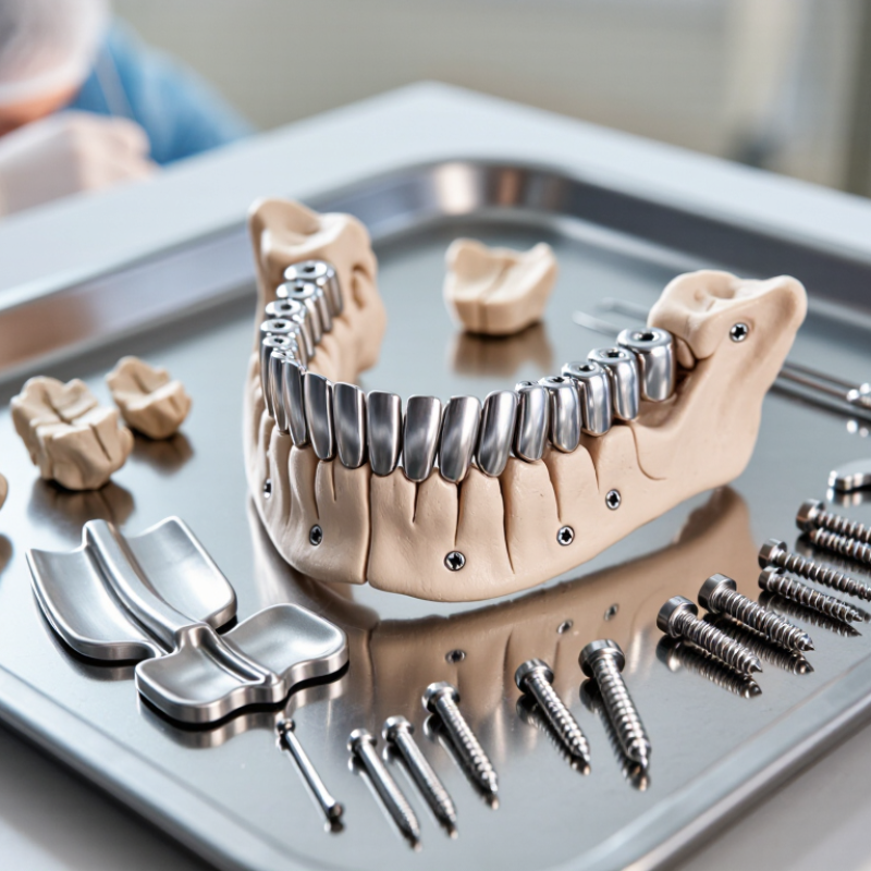
2026 Top Mandibular Reconstruction Plates Buying Guide

Mandibular Reconstruction Plates: Purpose, Structure, and Clinical Role

Mandibular Reconstruction Plates: Purpose, Structure, and Clinical Role

Mandibular reconstruction plates restore continuity after tumor removal, trauma, infection, or severe bone loss. The World Health Organization reported in 2022 that oral diseases affect nearly 3.5 billion people worldwide. GLOBOCAN 2022 recorded about 389,800 new lip and oral cavity cancer cases globally. These figures show why reliable reconstruction planning remains clinically important.

A plate usually includes a contoured body, screw holes, and fixation zones. Locking holes can stabilize screws when bone quality is reduced. The design must resist bending, torsion, and repeated chewing forces. Surgeons also consider plate thickness, curvature, defect length, and soft-tissue coverage. A small mismatch matters. Poor adaptation may increase stress around screws or disturb the intended occlusion.

Clinical experience shows that the “strongest” plate is not always the best choice. Patient anatomy, remaining bone, flap design, and postoperative function must guide selection. The AO Surgery Reference emphasizes stable fixation and accurate mandibular alignment as core reconstruction principles. Yet plate performance depends on more than hardware. Infection, inadequate coverage, and delayed healing can still compromise results. This is where buying decisions become less certain. Product data may describe mechanical strength, but clinical judgment must assess the whole patient. Even careful planning can require intraoperative adjustment.

Key Plate Types and Materials Used in Mandibular Reconstruction

Mandibular Reconstruction Plates: Key Types and Materials

Mandibular reconstruction plates restore continuity after trauma, tumor removal, or major bone loss. Plate selection depends on defect length, bone quality, occlusion, and soft-tissue coverage. In clinical planning, load-bearing plates often support the entire mandibular arch. Reconstruction plates are thicker and stronger than routine miniplates. Locking screw systems can improve stability when bone contact is limited. Non-locking plates remain useful when close adaptation is achievable. Curved, straight, and patient-specific designs address different mandibular shapes.

Titanium and titanium alloys are widely used because they combine strength, corrosion resistance, and relatively low weight. Their compatibility with imaging and biological tissues also supports postoperative assessment. Stainless steel may offer high strength, but it is less common in contemporary mandibular reconstruction. Material choice still requires careful review of allergies, imaging needs, and surgical technique. The strongest plate is not automatically the best choice. Fit matters.

In practice, surgeons assess plate thickness, hole configuration, screw direction, and bending requirements before fixation. A plate that is over-bent may lose mechanical reliability. Excessive contouring can create stress points. Poor soft-tissue coverage may also increase exposure risk. These details are easy to underestimate. I have found that preoperative virtual planning improves consistency, but it cannot replace intraoperative judgment. Anatomy rarely follows the plan perfectly. Product specifications, validated testing, and surgeon experience should guide final selection. Regional regulations and institutional protocols must also be checked before use.

How to Match Plate Design With Defect Location and Surgical Needs

Mandibular defects are not interchangeable. GLOBOCAN 2022 recorded 389,846 new lip and oral cavity cancer cases worldwide, creating highly varied reconstruction demands. A lateral body defect may need a pre-bent plate with strong load sharing. An angle defect needs accurate contour control near the ramus. Symphysis defects demand stable curvature and careful management of the mental nerves.

Plate thickness, screw position, and hole distribution should match bone quality and defect length. Longer spans may require stronger fixation and more bicortical purchase when safely possible. In contrast, thinner designs can reduce soft-tissue irritation in well-covered areas. A 2023 systematic review reported mandibular reconstruction plate exposure rates commonly near 20%, although techniques and follow-up differed between studies. That number should encourage caution, not automatic rejection.

Clinical experience still matters. A digitally planned plate can improve fit, but it cannot correct poor flap coverage or unstable occlusion. Surgeons should compare defect geometry, planned loading, vessel access, and soft-tissue thickness before selection. Small details matter. A misplaced hole can complicate fixation. A plate that looks ideal on a screen may feel excessive during exposure. These decisions are best documented with imaging, operative findings, and validated planning protocols, consistent with AO CMF guidance and applicable regulatory requirements.

2026 Top Mandibular Reconstruction Plates Buying Guide - How to Match Plate Design With Defect Location and Surgical Needs

Defect Location or Reconstruction Scenario Typical Biomechanical Demand Recommended Plate Design Typical Screw Strategy Key Surgical Requirements Main Selection Risks
Small lateral body defect
Continuity preserved; limited bone loss
Moderate functional loading with relatively short lever arms Low-profile or standard reconstruction plate with adequate contourability; compression or bridging design selected according to fracture and graft stability Bicortical fixation where anatomy permits; locking holes may help maintain fixation in compromised bone Accurate adaptation to the inferior border; avoid tooth roots, the mandibular canal, and the mental foramen An excessively bulky plate may irritate soft tissue; an undersized plate may lack fatigue resistance
Symphysis or anterior mandibular defect
Includes the chin and parasymphysis region
Multidirectional forces from mastication, including torsion and bending Rigid load-bearing reconstruction plate with a broad, anatomically contoured body; a design that resists torsion is preferred for larger defects Multiple fixation points on both sides of the defect; bicortical screws are commonly considered when safe and feasible Preserve mandibular width and chin projection; coordinate plate position with dental occlusion and the soft-tissue envelope Poor contouring can alter facial symmetry or occlusion; screw placement requires careful avoidance of tooth roots and the mental nerves
Segmental defect of the body
Continuity defect caused by tumor resection, trauma, or osteomyelitis
High load-bearing demand with bending and torsional stresses across a gap Load-bearing reconstruction plate with high fatigue strength, sufficient vertical height, and a stable bridging span; pre-contoured or patient-specific design can improve fit Distributed fixation on each remaining segment; locking fixation can reduce dependence on intimate plate-to-bone compression, especially in grafted or weakened bone Plan fixation around the mandibular canal and graft position; verify occlusion, inferior-border alignment, and the planned vascular pedicle pathway Insufficient plate stiffness, excessive unsupported span, repeated intraoperative bending, and poor seating can increase fatigue or exposure risk
Angle or posterior body defect
Transition between body and ramus
Combined bending and torsional loading at a geometrically complex transition zone Strong pre-contoured plate with adequate angular conformity and screw-hole access on both the body and ramus sides Use fixation points that provide a stable triangular or distributed construct while avoiding the mandibular canal and the inferior alveolar neurovascular bundle Ensure the plate does not impinge on the masseteric or pterygomandibular soft tissues; assess the available ramus height before selecting screw length Limited access, inadequate contour at the angle, and excessively long screws can compromise nerves, teeth, or adjacent soft tissue
Ramus defect without condylar replacement Load transfer is influenced by the vertical ramus and remaining temporomandibular joint anatomy Contoured load-bearing plate with a narrow or anatomically shaped superior portion to accommodate restricted access and limited bone stock Short, accurately directed screws may be required in the ramus; locking holes can be useful when bicortical purchase is limited Confirm the relationship to the sigmoid notch, coronoid process, mandibular canal, and temporomandibular joint A plate that is too wide or too rigid for the available ramus may restrict placement or cause soft-tissue irritation
Condylar or high ramus reconstruction
Loss of the condyle or articular unit
Requires restoration of mandibular height, movement, and joint function rather than plate strength alone Specialized condylar reconstruction system, graft-based reconstruction, or custom joint solution; a conventional body plate alone is generally insufficient Fixation must match the selected condylar or ramus reconstruction and the available posterior bone stock Preoperative imaging and occlusal planning are essential; evaluate facial nerve exposure, joint position, ramus height, and range-of-motion goals Treating the area as a simple plate-bridging problem may lead to malocclusion, altered mandibular motion, or joint-related complications
Large composite defect with soft-tissue loss
Bone and mucosal or cutaneous reconstruction required
High mechanical demand combined with elevated exposure and infection risk Rigid reconstruction plate with smooth contours, reliable fatigue resistance, and compatibility with free-flap or vascularized bone reconstruction Plan fixation to preserve the vascular pedicle and avoid excessive compression of the graft; locking fixation may be advantageous in selected cases Coordinate plate geometry with flap design, inset, skin paddle, dental rehabilitation, and anticipated postoperative imaging Plate prominence, dead space, compromised coverage, and interference with the pedicle can increase the risk of exposure or infection
Atrophic or osteoporotic mandible
Reduced bone height or poor cortical quality
Reduced screw purchase and greater risk of local stress concentration Load-sharing or load-bearing design selected according to continuity and graft status; broader contact distribution and locking capability may improve construct stability Use sufficient fixation points without overloading thin cortices; screw length and trajectory must be confirmed with imaging Avoid excessive plate bending and preserve remaining bone; consider the risk of fracture during drilling and screw insertion Over-tightening, inadequate edge distance, and excessive reliance on a small number of screws may cause loosening or cortical failure
Immediate dental rehabilitation planned
Implants or prosthetic restoration expected
Mechanical stability must be coordinated with future implant position and occlusal loading Patient-specific or digitally planned plate that preserves the intended dental arch, graft volume, and implant corridors Position screw holes and trajectories away from planned implants and dental roots; use virtual planning or cutting guides when available Integrate CT data, occlusion, resection margins, flap design, graft orientation, and implant planning before surgery A plate selected without dental planning may obstruct implants, compromise prosthetic alignment, or require revision
Revision or infected reconstruction
Previous hardware failure, nonunion, or contaminated field
Mechanical instability combined with impaired biology and increased infection risk Strong load-bearing plate selected after debridement and assessment of remaining bone; design should permit stable fixation outside compromised areas Place fixation in healthy, viable bone when possible; locking fixation may help where compression across the plate-bone interface is undesirable Assess union status, bone quality, bacterial contamination, soft-tissue coverage, and the cause of previous failure Replacing hardware without correcting infection, instability, malocclusion, or poor coverage may result in repeat failure
Planning note: Plate selection should be confirmed by the maxillofacial or reconstructive surgical team using clinical examination, occlusal assessment, and appropriate CT or three-dimensional planning. The final choice depends on defect size, remaining bone quality, graft type, soft-tissue coverage, fixation access, and the patient’s functional requirements.

Essential Buying Criteria for Strength, Fit, Safety, and Compatibility

2026 Top Mandibular Reconstruction Plates Buying Guide

Strength starts with evidence, not thickness alone. Published clinical reviews commonly report mandibular plate complications, including infection, loosening, and fracture, in roughly 10–30% of cases. Rates vary by defect size, radiation exposure, and patient health. Therefore, buyers should request fatigue-test results, bending limits, and documented clinical outcomes. A plate that feels rigid may still concentrate stress around screw holes.

Fit deserves equal attention. A 2023 systematic review of computer-assisted mandibular reconstruction found that digital planning can improve osteotomy accuracy and postoperative symmetry, although results depend on scanning, printing, and surgical execution. Ask whether the plate supports virtual planning, patient-specific contouring, or intraoperative adjustment. Check screw angulation, hole spacing, edge profile, and access for soft-tissue coverage. Small mismatches become obvious at the mandibular angle.

Safety information should be specific. Verify material composition, sterilization instructions, MRI conditions, and biological safety testing under ISO 10993 principles. Confirm compatibility with the intended screw diameter, driver system, and fixation technique. Regulatory clearance matters, but it does not replace surgeon judgment. I would also review adverse-event data and published follow-up periods. Short follow-up can hide late loosening. That is easy to overlook. A practical weakness remains: laboratory strength data rarely predicts every patient outcome. Teams should document this uncertainty before purchasing.

2026 Top Mandibular Reconstruction Plates Buying Guide

Essential Buying Criteria for Strength, Fit, Safety, and Compatibility

Tensile-strength comparison of commonly specified implant-grade titanium materials based on minimum values defined in ASTM F67 and ASTM F136. Higher material strength can support resistance to deformation, but final plate selection must also consider patient anatomy, screw compatibility, contouring requirements, fatigue performance, sterilization, and applicable biocompatibility requirements.

Reference basis: ASTM F67 Grade 2 commercially pure titanium and ASTM F136 titanium alloy Ti-6Al-4V ELI. Material properties are minimum specification values and may vary with product design, processing, thickness, and heat treatment.

Evaluation, Procurement, and Maintenance of Reconstruction Plate Systems

2026 Top Mandibular Reconstruction Plates Buying Guide
Evaluation, Procurement, and Maintenance of Reconstruction Plate Systems

Choosing a mandibular reconstruction plate system requires more than comparing hole patterns or purchase prices. Evaluation begins with anatomy, defect size, bone quality, fixation access, and the planned reconstructive method. Experienced surgical teams should review preoperative imaging and trial-fit options using patient-specific measurements. Assess plate geometry, screw compatibility, contouring limits, fatigue resistance, and radiographic visibility. Ask for documented testing, material information, traceability, and validated cleaning instructions. Evidence matters. A polished catalogue cannot replace clinical judgment.

During procurement, involve surgeons, maxillofacial nurses, sterile-processing staff, and purchasing specialists. Request consistent specifications across sizes, clear lot records, and responsive technical support. Compare total cost, not only the invoice. Include instruments, training, storage, replacement parts, and inspection time. Supplier qualification should include quality documentation, complaint handling, delivery reliability, and change-notification procedures. Do not accept vague claims. One overlooked detail can delay a case. Yet procurement plans are rarely perfect; teams should revisit assumptions after real procedures and near misses.

Tips: Store plates in clean, protected compartments, and inspect them under bright light before use. Look for scratches, distortion, corrosion, damaged threads, or missing labels. Follow the validated reprocessing cycle exactly. Never improvise with unapproved chemicals or undocumented repairs. Record each device’s identifier, procedure, sterilization history, and disposal decision. Routine audits can reveal recurring handling errors. Small findings matter. Maintenance should be practical, documented, and shared with the entire care team.

FAQS

What is a mandibular reconstruction plate used for?

It restores jaw continuity after trauma, tumor removal, or major bone loss. It supports the mandibular arch.

How should surgeons choose the plate type?

Consider defect length, bone quality, bite alignment, and soft-tissue coverage. Fit matters.

When are locking plates useful?

Locking screws can improve stability when bone contact is limited. They may help with difficult fixation access.

When can non-locking plates be appropriate?

Non-locking plates remain useful when the plate can closely follow the bone surface. Close adaptation is essential.

Which materials are commonly used?

Titanium and titanium alloys offer strength, corrosion resistance, and relatively low weight. Material choice still needs individual review.

Is the strongest plate always the best choice?

No. Thickness, shape, screw direction, imaging needs, and tissue coverage also matter. More strength is not automatically better.

What problems can excessive contouring cause?

Over-bending may reduce mechanical reliability. Sharp bends can create stress points. Plans can be wrong.

What should a hospital evaluate before purchasing a system?

Review geometry, screw compatibility, fatigue testing, traceability, cleaning instructions, and radiographic visibility. A catalogue cannot replace evidence.

What costs should procurement teams consider?

Include instruments, training, storage, replacement parts, and inspection time. The invoice is only one part.

How should plates be inspected and maintained?

Inspect them under bright light for scratches, distortion, corrosion, damaged threads, or missing labels. Follow validated reprocessing instructions exactly. Record every device identifier and sterilization history.

Conclusion

This 2026 buying guide explains how Mandibular Reconstruction Plates support the restoration of jaw continuity, stability, and function after trauma, tumor removal, infection, or corrective surgery. It reviews the role, structure, and clinical applications of reconstruction plates, along with common plate designs and material options. Readers will learn how factors such as defect location, bone quality, load requirements, surgical access, and soft-tissue conditions influence the selection of straight, curved, locking, or patient-specific systems.

The guide also presents practical purchasing criteria, including mechanical strength, anatomical fit, corrosion resistance, imaging compatibility, fixation performance, and instrument compatibility. It outlines a careful evaluation and procurement process that considers documentation, sterilization requirements, inventory control, and surgical workflow. Finally, it highlights the importance of inspection, cleaning, storage, and maintenance procedures to help preserve system reliability and support safe, efficient clinical use.

Isabella

Isabella

Isabella is a dedicated marketing professional with a sharp focus on driving brand growth and engagement through strategic content creation. With an extensive background in digital marketing, she combines her passion for storytelling with her keen understanding of industry trends to deliver......