Bonevia
Choosing Cancellous Screws in 2026 requires more than selecting the largest diameter available. Bone quality, fracture geometry, thread purchase, and healing biology must work together. A screw that looks ideal on a product chart may fail in soft metaphyseal bone. Small differences matter, such as thread depth, pitch, head design, and washer compatibility.
Dr. Paul Tornetta III, a respected orthopedic trauma surgeon, has often stressed a practical principle: “Fix the fracture, but respect the biology.” This idea remains highly relevant when comparing Cancellous Screws. Stable compression can support healing, but excessive compression may damage fragile bone or reduce local blood supply. The correct choice depends on the fracture pattern and the patient, not on marketing language.
In this guide, we examine how surgeons can assess screw diameter, length, thread configuration, and material in 2026. Radiographs provide useful evidence. CT scans may reveal hidden comminution or poor bone stock. Intraoperative feedback also matters, especially when purchase feels unexpectedly weak. Manufacturer instructions, validated surgical techniques, and current clinical evidence should guide final selection.
No checklist is flawless.
Even experienced surgeons can misjudge depth by a few millimeters. That small error may affect joint clearance, fixation strength, or soft-tissue irritation. Therefore, this discussion includes practical limitations, common selection mistakes, and questions worth revisiting before implantation. The goal is not to promote one device. It is to support careful, evidence-informed decisions about Cancellous Screws, with patient safety and biological healing kept at the center.
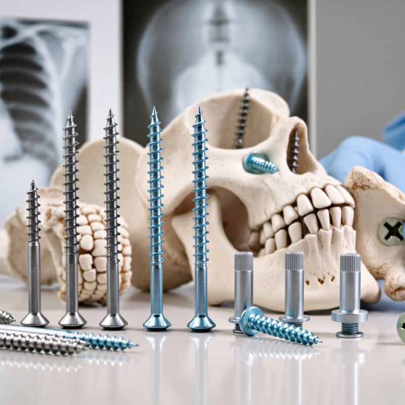
Cancellous screws are orthopedic implants designed to secure fragments in softer, porous bone. This bone is common near joints, including the metaphysis and epiphysis. Unlike cortical screws, they usually have deeper, wider threads and a larger thread pitch. These features improve purchase in cancellous bone, where the internal structure resembles a honeycomb. Simple image: threads grip the spaces.
Their basic function is stabilization. A cancellous screw can hold a fracture, attach a small bone fragment, or provide compression across a fracture line. In a lag technique, the threads engage the far fragment while the near hole allows controlled sliding. Tightening the screw draws the fragments together. A washer may help distribute pressure when the outer bone surface is weak or uneven.
Choosing the screw requires more than matching its diameter to the visible fracture. The surgeon considers bone quality, fragment size, fracture depth, thread length, and the required compression. A fully threaded design may provide positional fixation, while a partially threaded design may support lag compression. This distinction is easy to miss. It can affect stability.
Clinical imaging and direct assessment guide the final selection. Length should provide secure purchase without entering a joint or damaging nearby structures. The ideal screw is not always the longest one. In practice, anatomy can be less predictable than a radiograph suggests, so measured data and surgical judgment must work together. There is no universal choice.
How to Choose Cancellous Screws in 2026?
Key Factors for Matching Screw Design to Bone and Procedure
Cancellous screw selection begins with bone quality, fracture location, and the reduction goal. Metaphyseal bone is softer and has wider trabecular spaces than dense cortical bone. A larger thread profile can improve purchase without excessive insertion force. Measure twice. Screw diameter should fit the fragment and avoid splitting it. Length must provide adequate thread engagement while preventing joint penetration.
Thread design should match the intended function. A partially threaded screw can create interfragmentary compression when its threads remain beyond the near fragment. A fully threaded option may better preserve alignment or support controlled fixation. Thread length, pitch, and core diameter affect purchase and insertion torque. A washer can distribute load over fragile outer bone. It may also irritate soft tissue if placed carelessly.
Procedure details change the decision. Fluoroscopy or equivalent imaging should confirm trajectory, depth, and joint safety. Preoperative scans can reveal poor bone stock, hidden comminution, or an unusual anatomy. In my experience, selecting the longest screw is not automatically safer. Excessive length can damage cartilage or nearby structures. Surgical training, validated technique guidance, and the device’s instructions remain essential. Even a careful checklist has limits; patient movement, imperfect reduction, and changing bone density can alter the final choice. Documenting these uncertainties improves follow-up decisions.
| Selection Dimension | Typical Options or Data | Best-Matched Bone or Procedure | Why It Matters | Key Verification Point |
|---|---|---|---|---|
| Thread Configuration |
Partially threaded screws have a smooth shaft and threaded distal portion. Fully threaded screws engage bone along the threaded portion. |
Partially threaded: lag fixation and interfragmentary compression. Fully threaded: positional fixation, comminuted fragments, or situations where compression is not desired. |
A partially threaded screw can achieve compression when the near cortex is overdrilled and the far fragment is securely engaged. A fully threaded screw generally provides more uniform purchase but does not automatically create lag compression. | Confirm that the thread length is sufficient to cross the fracture while allowing the threads to obtain purchase in the far fragment. |
| Thread Pitch and Profile | Cancellous screws usually have a larger pitch and deeper thread profile than cortical screws. Common systems provide coarse cancellous threads designed for trabecular bone. | Metaphyseal and epiphyseal bone, where trabecular structure is less dense than diaphyseal cortical bone. | Coarse, deep threads can improve purchase in cancellous bone by increasing engagement with the trabecular architecture and distributing load over a larger area. | Avoid assuming that a coarse thread compensates for poor placement, insufficient depth, or severely deficient bone stock. |
| Core Diameter | The core is smaller than the major thread diameter. A larger core generally improves bending and torsional strength, while a smaller core may offer more thread depth relative to the shaft. |
Larger core: higher mechanical demand or longer working length. Smaller core: smaller fragments or limited bone volume, when adequate strength remains. |
Core diameter affects fatigue resistance, insertion torque, and the amount of bone preserved around the screw. | Select the smallest diameter that provides adequate stability without causing fragment splitting or loss of purchase. |
| Nominal Diameter | Common cancellous screw systems include small-fragment and large-fragment ranges; exact diameters vary by fixation system and anatomy. |
Smaller diameters: small fragments, pediatric or anatomically constrained areas. Larger diameters: larger metaphyseal fragments and higher-load fixation. |
Diameter influences pull-out resistance, bending strength, insertion torque, and the risk of weakening the fragment. | Match the screw diameter to the available bone corridor and the associated drill, tap, guide, and instrument system. |
| Screw Length | Choose a length that places the threaded portion fully within the intended purchase fragment without unnecessary penetration beyond the far cortex. |
Lag fixation: threads should normally engage the far fragment rather than the near fragment. Positional fixation: threads should engage the planned bone segments according to the construct. |
Excessive length may irritate soft tissues or endanger nearby structures. Insufficient length reduces thread engagement and fixation security. | Measure along the prepared trajectory and account for countersinking, washers, oblique placement, and the implant system’s measurement method. |
| Head Design | Options include hexagonal or other drive recesses, low-profile heads, and heads designed for use with washers or plates. |
Low-profile head: areas with limited soft-tissue coverage. Washer-compatible head: weak metaphyseal cortex or larger load-bearing contact area. |
Head geometry affects insertion control, stripping risk, soft-tissue prominence, and load transfer at the near cortex. | Ensure the driver is fully seated and compatible with the screw recess. Do not use a damaged driver or a mismatched instrument. |
| Washer Use | A washer increases the contact area under the screw head and can reduce the risk of the head sinking into weak or thin cortex. | Osteoporotic metaphyseal bone, thin cortical shell, oblique screw placement, or fixation near a joint surface. | A washer can spread compressive force over a wider area and help prevent localized cortical indentation. | Confirm soft-tissue clearance and compatibility with the screw head. A washer does not replace correct trajectory or adequate thread purchase. |
| Cannulated vs. Solid | Cannulated screws are inserted over a guidewire. Solid screws are inserted without a central guidewire channel. |
Cannulated: precise trajectory, minimally invasive placement, or proximity to a joint surface. Solid: straightforward exposure, higher resistance to guidewire-related complications, or when cannulation is unnecessary. |
Guidewire-assisted placement can improve targeting and repeatability, while solid screws may simplify instrumentation and eliminate central-channel concerns. | Check guidewire position repeatedly and prevent guidewire advancement beyond the intended depth or into critical structures. |
| Material | Common implant materials include stainless steel and titanium alloys. Material properties differ in elastic modulus, strength, imaging behavior, and compatibility with other implants. | Titanium alloy may be considered when reduced radiographic artifact or compatibility with a titanium construct is important. Stainless steel may be selected where high strength and system compatibility are priorities. | Material affects stiffness, corrosion considerations, artifact on imaging, and the behavior of mixed-metal constructs. | Follow the implant system instructions regarding material pairing, MRI conditions, and compatibility with plates, washers, or other implants. |
| Bone Quality | Assess trabecular density, cortical thickness, comminution, bone loss, osteoporosis, and the quality of the available purchase zone. | Poor bone quality may require a longer thread engagement zone, a larger contact area, supplemental fixation, a washer, or an alternative construct. | Screw pull-out resistance depends strongly on bone density, insertion depth, thread engagement, and the direction and magnitude of applied load. | Do not rely on screw diameter alone to compensate for deficient bone. Evaluate the complete fixation strategy. |
| Joint-Surface Clearance | The screw tip and any guidewire must remain outside the articular surface unless the procedure specifically requires otherwise. | Epiphyseal fractures, periarticular fixation, and procedures involving the femoral head, tibial plateau, ankle, wrist, or other joint-adjacent anatomy. | Even small errors in trajectory or length can cause cartilage injury, hardware prominence, or restricted joint motion. | Use orthogonal or multiplanar imaging as appropriate and verify final screw position before completing fixation. |
| Compression Requirement | Compression may be created by lag technique, eccentric placement in a plate, or a dedicated compression feature in a compatible system. | Anatomical fracture reduction, osteotomy fixation, and selected arthrodesis procedures where controlled interfragmentary compression is desired. | Compression can improve fragment contact and construct stability, but excessive compression may compromise blood supply or displace small fragments. | Confirm reduction before tightening and apply compression gradually while monitoring fragment position and soft-tissue conditions. |
| Insertion Technique | Typical steps may include drilling, measuring, optional tapping according to bone quality and system design, countersinking when indicated, and controlled insertion. | All cancellous screw procedures, particularly when using lag fixation or when the near cortex is dense. | Correct preparation reduces insertion torque, improves seating, limits fragment rotation, and lowers the risk of drive recess damage. | Use only the specified drill, tap, countersink, guidewire, and driver. Follow the current surgical technique and implant instructions. |
| Construct and Load Sharing | Evaluate whether the screw will be used alone, with a washer, or as part of a plate, tension-band, or multifragmentary fixation construct. | High-load regions, unstable fractures, comminuted patterns, and osteoporotic bone often require load sharing across more than one fixation element. | A single screw may provide compression but may not adequately resist rotation, bending, or shear without additional fixation. | Match the screw to the fracture pattern and the overall construct rather than selecting it in isolation. |
| Postoperative Imaging and MRI | Radiographic visibility, artifact, and MRI conditions depend on implant material, geometry, and the complete implant system. | Patients likely to need postoperative CT, MRI, or long-term radiographic monitoring. | Imaging compatibility affects follow-up assessment, fracture-healing evaluation, and detection of complications. | Review the current device labeling for MRI safety, scan conditions, artifact expectations, and any restrictions on mixed implants. |
How to Choose Cancellous Screws in 2026?
Choosing a cancellous screw begins with bone quality, fracture geometry, and the required compression. The diameter should provide strength without splitting the cancellous envelope. In osteoporotic bone, a larger diameter may improve purchase, but excessive drilling can weaken the fragment. AO Surgery Reference advises matching screw size to fragment dimensions and the available safe corridor. That judgment remains practical, not automatic.
Length deserves equal attention. Measure the planned trajectory, then subtract the depth needed to avoid joint penetration. A screw should cross the fracture and obtain sufficient thread purchase in the far fragment. Too short, and fixation may loosen. Too long, and the tip can damage cartilage or soft tissue. The 2024 National Joint Registry report tracked more than four million hip and knee procedures, reminding clinicians how frequently bone quality and fixation decisions affect older patients. However, its data does not directly validate one screw length for every fracture.
Thread pattern changes the mechanical goal. A partially threaded screw can create interfragmentary compression when the threads remain beyond the fracture line. A fully threaded design may better maintain alignment or control a short fragment, but it will not compress automatically. Check the thread pitch, available purchase, and reduction before insertion. Do not trust the catalog chart alone. Real anatomy is less cooperative. Cadaveric and clinical evidence also varies, so the final choice should be reviewed against current AO and OTA guidance, imaging, and the patient’s bone quality.
Common cancellous screw systems include nominal diameters around 4.0–4.5 mm for smaller fragments and 6.5–7.3 mm for larger fragments or higher fixation demands. The correct diameter should preserve bone stock while providing adequate purchase.
Select length by measuring the available bone corridor and confirming screw position with imaging. Use partially threaded screws when interfragmentary compression is required; use fully threaded screws when positional fixation or consistent thread engagement is the priority. Always follow the applicable surgical technique and device instructions.
Material, coating, and cannulation shape how a cancellous screw performs in real clinical conditions. Stainless steel offers high strength and familiar handling. Titanium alloy is lighter and creates fewer imaging artifacts. However, titanium may provide different bending behavior, so the surgeon must match stiffness with the fracture pattern and bone quality. Osteoporotic metaphyseal bone needs careful thread engagement, not simply a stronger screw.
Coatings deserve closer scrutiny. A porous or osteoconductive surface may support bone attachment, but its value depends on coating adhesion, surface design, and clinical evidence. A coating cannot compensate for poor reduction or inaccurate screw placement. Check the technical documentation, sterilization instructions, and applicable quality standards. Small surface defects can matter. They may be overlooked during a busy procedure.
Cannulated screws can guide placement over a wire, which helps when access is limited or alignment is difficult. The wire must remain stable. The cannula, drill, and screw should be compatible. Blood or bone debris can obstruct the channel. That risk is easy to underestimate. Intraoperative imaging also deserves attention, especially near joints. I would compare the complete system rather than the screw alone. The most expensive option is not automatically the safest, and no material or coating suits every patient.
Safe cancellous screw selection begins with the fracture pattern, bone quality, and intended load. The surgeon should confirm screw diameter, length, thread design, and material compatibility with current imaging. A preoperative radiograph or CT scan can reveal weak purchase, joint involvement, or hidden comminution. Small details matter. The screw should cross the target fragment without entering the joint. Depth measurement must include the planned trajectory, not guesswork. Intraoperative imaging can confirm position in more than one view. One view may deceive.
Placement checks should include the entry point, insertion angle, and final tip location. The surgeon should avoid excessive torque, which may strip soft cancellous bone. A washer can help distribute force when the cortex is thin, but it is not suitable for every fracture. Fixation should be tested gently, without destabilizing the repair. A tight screw is not automatically a stable screw. Poor bone quality may require a different construct or additional fixation. This is where judgment matters, and even experienced teams should review assumptions.
Postoperative care deserves equal attention. Record circulation, sensation, movement, swelling, and skin condition before discharge. Give clear instructions about weight bearing, elevation, wound care, and warning signs. Increasing pain, drainage, fever, numbness, or a cold digit needs prompt medical assessment. Follow-up imaging should confirm maintained alignment and progressive healing. I would also question routine schedules when healing is delayed. Patient age, smoking status, nutrition, medication use, and activity level can change the plan. The final choice should follow current clinical guidance and the treating surgeon’s examination.
Consider bone quality, fracture location, fragment size, and the intended fixation goal. Soft metaphyseal bone may need broader threads for better purchase. Measure carefully. Diameter and length must fit without splitting the fragment or entering the joint.
Its threads can remain beyond the near fragment and create interfragmentary compression. This may help close a fracture gap. The technique depends on accurate reduction and suitable thread length. Compression is not guaranteed when positioning is imperfect.
A fully threaded screw may preserve alignment or provide controlled fixation. It can suit situations where compression is not the main goal. Thread pitch, core diameter, and insertion torque still matter. More threads do not automatically mean better stability.
Measure along the planned trajectory, not a straight surface distance. The screw should engage enough bone without reaching cartilage or nearby structures. The longest option is not automatically safer. One extra millimeter can matter near a joint.
Stainless steel offers familiar handling and high strength. Titanium alloy is lighter and may create fewer imaging artifacts. Its bending behavior differs, so stiffness must match the fracture pattern. A stronger screw cannot correct weak bone purchase.
Some porous or bone-conductive surfaces may support bone attachment. Their value depends on adhesion, surface design, and clinical evidence. A coating cannot correct poor reduction or inaccurate placement. Small surface defects deserve attention.
The guidewire must remain stable during drilling and insertion. The wire, drill, cannula, and screw should be compatible. Blood or bone debris can block the channel. That risk is easy to underestimate. Imaging should confirm the trajectory and final tip position.
Confirm the entry point, insertion angle, depth, and tip location in multiple views. Avoid excessive torque, which can strip soft cancellous bone. A tight screw is not always a stable screw. After surgery, monitor circulation, sensation, movement, swelling, and skin condition. Increasing pain, drainage, fever, numbness, or a cold digit requires prompt medical assessment.
Choosing Cancellous Screws in 2026 requires a clear understanding of their purpose, design, and interaction with softer, porous bone. These screws are commonly used to achieve stable fixation in areas where bone structure differs from dense cortical regions. Selection should begin with the procedure, anatomical location, bone quality, and required holding strength. Diameter and length must provide sufficient purchase without damaging surrounding tissue, while thread depth and pitch should match the bone and desired compression or stabilization effect.
Material, surface coating, and cannulation are additional factors to compare, especially when imaging guidance, insertion control, or long-term compatibility is important. Before placement, clinicians should confirm screw trajectory, depth, alignment, and the absence of interference with joints, nerves, or blood vessels. Postoperative care should include monitoring for loosening, infection, poor healing, or loss of fixation. Final decisions should follow current clinical protocols, imaging findings, and the judgment of a qualified orthopedic professional.