Bonevia
Choosing the right Acutrak Screws is not a simple matter of matching a product name to a fracture. Screw selection can influence compression, stability, healing conditions, and the surgeon’s ability to protect surrounding tissues. A reliable decision begins with the patient’s anatomy, the fracture pattern, bone quality, and the intended fixation strategy.
The Acutrak system includes different screw designs, sizes, and thread configurations. Each option may suit a particular surgical situation. Length matters. Diameter matters. Even the entry point matters. A screw that appears suitable on a radiograph may be unsuitable after considering cortical thickness, joint proximity, or the available purchase in small bone. Surgeons should review current manufacturer instructions, imaging, and relevant institutional protocols before choosing an implant.
Small details can change the plan.
This guide will examine practical factors behind Acutrak Screws selection, including compression requirements, implant dimensions, bone density, and surgical access. It will also consider common judgment errors, such as relying on a familiar size or treating every fracture pattern alike. A neat sizing chart cannot replace clinical assessment. That limitation deserves attention. Product availability, surgeon training, and patient-specific risks may also influence the final decision. The information here supports informed discussion, but it does not replace a qualified orthopedic surgeon’s evaluation, operative planning, or the manufacturer’s official guidance.
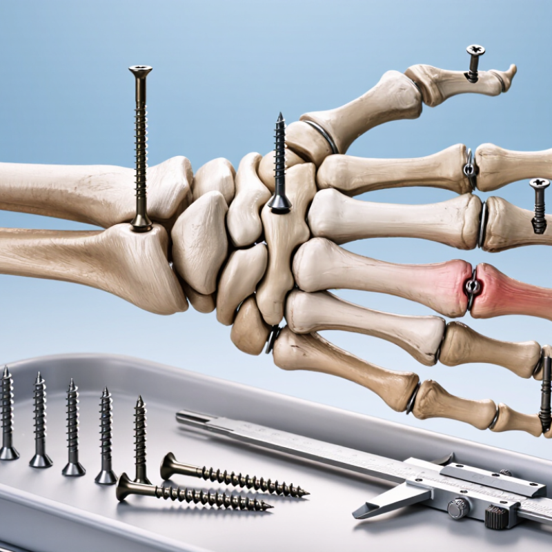
Headless compression screws use changing thread pitches to draw two bone fragments together as the screw advances. Their buried profile can reduce soft-tissue irritation near joints. Cannulation also supports guidewire placement and more controlled trajectories. However, compression is not automatic. Poor reduction, weak bone, or an incorrect entry point can undermine the design.
Clinical selection should match anatomy and loading demands. Small-diameter screws may suit phalangeal, carpal, or osteochondral fixation. Larger diameters may offer stronger purchase in selected fractures, but they remove more bone. A 2023 review in the Journal of Orthopaedic Trauma reported high union rates for headless compression screw fixation in small-joint injuries, while also noting complications such as migration and loss of compression. Numbers vary widely by anatomy and study design. That matters.
Market data provides context, not surgical guidance. Grand View Research estimated the global orthopedic screws market at more than USD 1 billion in 2023, with continued growth expected through the decade. This expansion reflects broader use, not proof that every screw fits every fracture. In practice, surgeons assess fracture geometry, bone quality, screw length, thread engagement, and fluoroscopic views. A sizing chart cannot replace that judgment. I would also question overly confident claims about “optimal” compression, because cadaveric performance may differ from healing bone. Thin fragments remain unforgiving.
Choosing the right headless compression screw starts with the fracture, not the implant tray. A simple, stable fracture may need one screw with controlled compression. A long oblique or rotationally unstable fracture may require additional fixation. Comminution changes the plan again. Small fragments can split under excessive compression.
The anatomy demands equal attention. Measure the available bone length and identify nearby joints, tendons, nerves, and vessels. In a narrow phalanx, a large screw may weaken the cortex or irritate soft tissue. In cancellous bone, thread placement and purchase can affect stability. The screw should cross the fracture securely without entering the joint. That sounds obvious. It is still missed.
Fixation requirements depend on load, bone quality, and patient activity. A screw must provide compression while resisting rotation and shear. Poor bone may need a longer thread segment, a different trajectory, or supplemental fixation. Imaging should confirm fracture reduction before final insertion. Intraoperative judgment remains essential, especially when the fracture pattern differs from preoperative scans. A useful plan should include alternatives, because anatomy rarely follows the drawing perfectly. My clinical reasoning would also question whether compression is appropriate at all; some fractures need alignment and protection more than aggressive compression. Surgical decisions should follow validated techniques, current evidence, and the treating specialist’s assessment.
| Fracture or Clinical Scenario | Typical Anatomical Sites | Key Anatomical Assessment | Recommended Screw Characteristics | Fixation Requirements | Important Planning Considerations |
|---|---|---|---|---|---|
| Simple transverse or short-oblique fracture | Phalanges, metacarpals, metatarsals, small periarticular fragments | Fragment length, cortical thickness, available bone stock, and proximity to the joint surface | Headless compression screw with a diameter appropriate to the bone and a length sufficient to obtain bicortical or secure fragment purchase | Compression and rotational control | Place the screw as close as practical to perpendicular to the fracture plane. Confirm that the screw will not enter the joint or damage nearby neurovascular structures. |
| Long-oblique or spiral fracture | Metacarpals, metatarsals, phalanges, distal radius, and other long-bone segments | Fracture-line length, risk of shortening, and whether one or more fixation points are needed | Longer screw or multiple screws may be required; screw diameter should preserve adequate surrounding cortex | Axial compression plus anti-rotation | A single screw may not adequately control rotation. Consider additional fixation when the fracture is long, unstable, or associated with comminution. |
| Intra-articular fracture | Carpal bones, phalangeal condyles, radial styloid, and other joint-involving fragments | Articular surface congruity, fragment size, subchondral bone thickness, and safe screw trajectory | Low-profile or fully buried headless screw with a diameter that fits the fragment without splitting it | Anatomic reduction and compression | All components of the screw must remain below the articular surface. Fluoroscopy or other imaging should be used to verify reduction, trajectory, and final depth. |
| Small osteochondral or marginal fragment | Joint margins, carpal bones, radial or ulnar styloid fragments | Fragment dimensions, cartilage condition, subchondral support, and risk of fragment fragmentation | Small-diameter headless screw selected to maximize purchase while minimizing bone removal | Preserve fragment integrity | A screw that is too large can split the fragment or compromise cartilage. In very small or poorly supported fragments, alternative fixation may be more appropriate. |
| Small-bone fracture with limited bone stock | Scaphoid, lunate, phalanges, metacarpals, and other compact bones | Bone width, screw-axis length, vascularity, and available purchase on both sides of the fracture | Smallest suitable screw diameter with adequate thread engagement and a length that avoids overpenetration | Stable fixation with minimal bone sacrifice | Use precise imaging and a guidewire when appropriate. Avoid violating adjacent joints, tendon paths, and critical vascular structures. |
| Comminuted fracture | Distal radius, metacarpals, phalanges, and periarticular regions | Number and size of fragments, soft-tissue condition, bone loss, and ability to reconstruct the joint surface | Screw selection should be based on the largest stable fragments; a headless screw may be combined with other fixation methods | Supplemental stability may be necessary | Headless compression screws alone may not control multiple fragments or restore length. Avoid excessive compression that could collapse or devascularize small fragments. |
| Nonunion or delayed union requiring compression | Scaphoid, phalanges, metacarpals, and selected periarticular bones | Nonunion type, bone viability, deformity, gap size, sclerosis, and presence of infection | Compression screw with sufficient length and thread purchase; bone grafting or corrective osteotomy may be required | Compression plus biological restoration | Correct deformity and prepare the nonunion site as indicated. Assess vascularity and exclude infection before definitive fixation. |
| Osteoporotic or low-density bone | Older patients, metaphyseal regions, and bones affected by reduced mineral density | Bone quality, cortical support, fragment stability, and risk of screw loosening or cutout | Choose a diameter and length that maximize available purchase without compromising the cortex or joint | Purchase may be limited | Compression should be controlled to prevent fragment collapse. Supplemental fixation or a different construct may be needed when screw purchase is inadequate. |
| Avulsion fracture or ligament-related fragment | Collateral ligament attachments, tendon insertions, and small periarticular avulsion sites | Fragment size, soft-tissue attachment, joint stability, and direction of traction forces | Small headless screw only when the fragment can safely accept the implant; alternative anchors or wires may be considered | Restore soft-tissue stability | Do not compromise the attached ligament or tendon. The fixation method should address both fragment stability and the associated soft-tissue injury. |
| Fracture requiring buried hardware | Subcutaneous bones, articular regions, and areas exposed to tendon or footwear pressure | Soft-tissue coverage, tendon glide, implant prominence risk, and required depth below the surface | Headless design with fully countersunk or buried placement, provided adequate fixation strength is maintained | Minimize hardware irritation | Verify that the implant is fully below the bone surface without excessive countersinking, which may weaken the fragment or reduce purchase. |
Choosing the right headless compression screw starts with anatomy, not the catalog. Small screws suit narrow phalanges and limited bone stock. Larger diameters can improve purchase, but they remove more bone and may create a stress concentration. AO Surgery Reference guidance stresses matching diameter to fragment size, cortical thickness, and fracture pattern.
Thread design changes compression behavior. A partially threaded screw can create lag compression when the near cortex is overdrilled. A fully threaded option may preserve alignment better, but it usually needs a separate compression strategy. Differential-pitch threads generate compression as the screw advances. That can be useful in a short oblique fracture. It can also over-compress a fragile fragment. Be careful here.
The 2024 Grand View Research market analysis estimated the global orthopedic screws market at approximately US$1.1 billion in 2023, reflecting broad use across trauma and reconstruction. That figure does not prove one design is clinically superior. A 2023 review in Foot and Ankle Orthopaedics likewise emphasized that fixation strength depends on bone quality, screw trajectory, and construct stability, not diameter alone. In practice, measure the planned path, check the far cortex, and confirm that the threads cross the intended fragment. My checklist still feels incomplete when bone is osteoporotic or fragments are tiny. Surgeon judgment remains essential.
Screw diameter should match the fragment size, anatomy, and required fixation strength. Smaller screws, such as 2.5 mm, are commonly selected for small bones and limited fragments, while larger 4.0–5.5 mm screws may provide greater structural strength where bone volume permits. Variable-pitch threads create compression as the screw advances, whereas partially threaded designs can provide lag compression when the near fragment is appropriately prepared. Final selection should be based on imaging, bone quality, fracture pattern, and the implant system’s surgical technique.
Choosing the right headless compression screw begins with the patient, not the implant tray. Review fracture location, bone quality, soft-tissue condition, activity level, and healing risks. A small, stable fracture in dense bone may need a different screw diameter than a comminuted fracture in osteoporotic bone. Check radiographs carefully. Use advanced imaging when fracture geometry remains uncertain.
Surgical Technique Surgical technique strongly influences fixation. Establish the guidewire trajectory before drilling. Keep it centered on both anteroposterior and lateral views. Measure screw length cautiously, because a few extra millimeters can irritate cartilage or soft tissue. Prepare the entry site according to the system’s instructions. Compression should close the fracture without crushing fragile fragments. Gentle handling matters.
Instrumentation Instrumentation must support accuracy under pressure. Confirm that the guidewire, drill, depth gauge, and screwdriver work together before incision. A loose connection can redirect the wire. Poor fluoroscopic positioning can also create false confidence. It happens. Keep backup instruments available, especially when the first guidewire path is unsatisfactory. The selected screw should match the prepared channel and the intended compression effect.
In Practice In practice, a neat preoperative plan may still change after exposure. Unexpected bone loss, unstable fragments, or limited access can alter the safest option. Reassess rather than forcing the original plan. Document the reasoning, imaging findings, and final construct. Evidence and training guide the decision, but careful observation during surgery remains essential.
Selecting the appropriate headless compression screw begins with the procedure, not the product name.
Match the implant to the fracture pattern, bone size, and required compression. A small articular fragment may need a narrow screw with controlled insertion. A longer fracture line may require greater thread engagement and a carefully chosen trajectory. The screw should support reduction without crossing vulnerable joints or soft tissues.
Preoperative imaging is essential. Review multiple views, and consider three-dimensional imaging when the fracture is complex. Measure the intended path, then allow enough room for the screw head to sit beneath the surface. Thread length matters. Too much threaded area can block compression, while too little may reduce purchase. Bone quality also changes the plan. Osteoporotic bone may require a larger diameter, a different fixation strategy, or additional stabilization.
Small details matter.
Confirm the guidewire position before drilling. Check the screw length twice. A measurement can still be wrong. Intraoperative imaging should verify depth, alignment, and joint clearance before final seating. Experienced surgeons also consider swelling, tissue tension, and the patient’s expected loading demands. The ideal choice is not always the longest or strongest option. It is the implant that fits the anatomy, preserves reduction, and supports safe healing. Mistakes often begin when selection follows habit instead of the actual procedure.
Start with the fracture pattern, not the implant tray. Stable fractures may need one screw, while unstable fractures may require additional fixation.
Long oblique or rotationally unstable fractures need careful control. Comminuted fragments can split when compression becomes excessive.
Measure bone length and locate nearby joints, tendons, nerves, and vessels. The screw must cross the fracture securely without entering the joint.
Not always. In a narrow phalanx, a large screw may weaken the cortex or irritate soft tissue. Smaller may be safer.
Consider load, bone quality, fracture stability, and patient activity. Poor bone may need a longer thread segment or supplemental fixation.
Measure cautiously and confirm the available bone length. A few extra millimeters can irritate cartilage or soft tissue.
Establish the trajectory before drilling. Keep the wire centered on anteroposterior and lateral imaging views.
Confirm that the guidewire, drill, depth gauge, and screwdriver work together. Keep backup instruments available. Equipment can fail.
Reassess after exposure if bone loss, unstable fragments, or limited access appears. Forcing the first plan may create avoidable problems.
No. Some fractures need alignment and protection more than aggressive compression. This judgment should follow imaging, evidence, training, and specialist assessment.
Choosing the right Acutrak Screws requires a structured evaluation of fracture characteristics, patient anatomy, and the intended goals of fixation. Their headless, compression-oriented design can support stable fracture alignment while minimizing hardware prominence, but selection should be based on the location, size, pattern, and stability of the fracture. Surgeons should consider whether the procedure requires focused compression, interfragmentary stability, or a combination of fixation strategies.
Important factors include screw diameter, length, thread configuration, and available compression options. Patient bone quality, soft-tissue condition, healing potential, and relevant medical history should also influence the decision. Proper surgical technique, accurate measurement, drilling, insertion, and use of compatible instrumentation are essential for reliable placement. Ultimately, the most appropriate Acutrak Screws are selected by matching their mechanical properties and dimensions to the anatomy and clinical requirements of each procedure, while prioritizing safe handling, secure fixation, and effective postoperative recovery.